The top two bones of the neck, C1 (the atlas) and C2 (the axis), form a joint that provides about half of your ability to turn your head. When this joint wears out with arthritis, it can cause intense pain at the base of the skull and up the back of the head. The nearby greater occipital nerve can become irritated, causing occipital neuralgia.
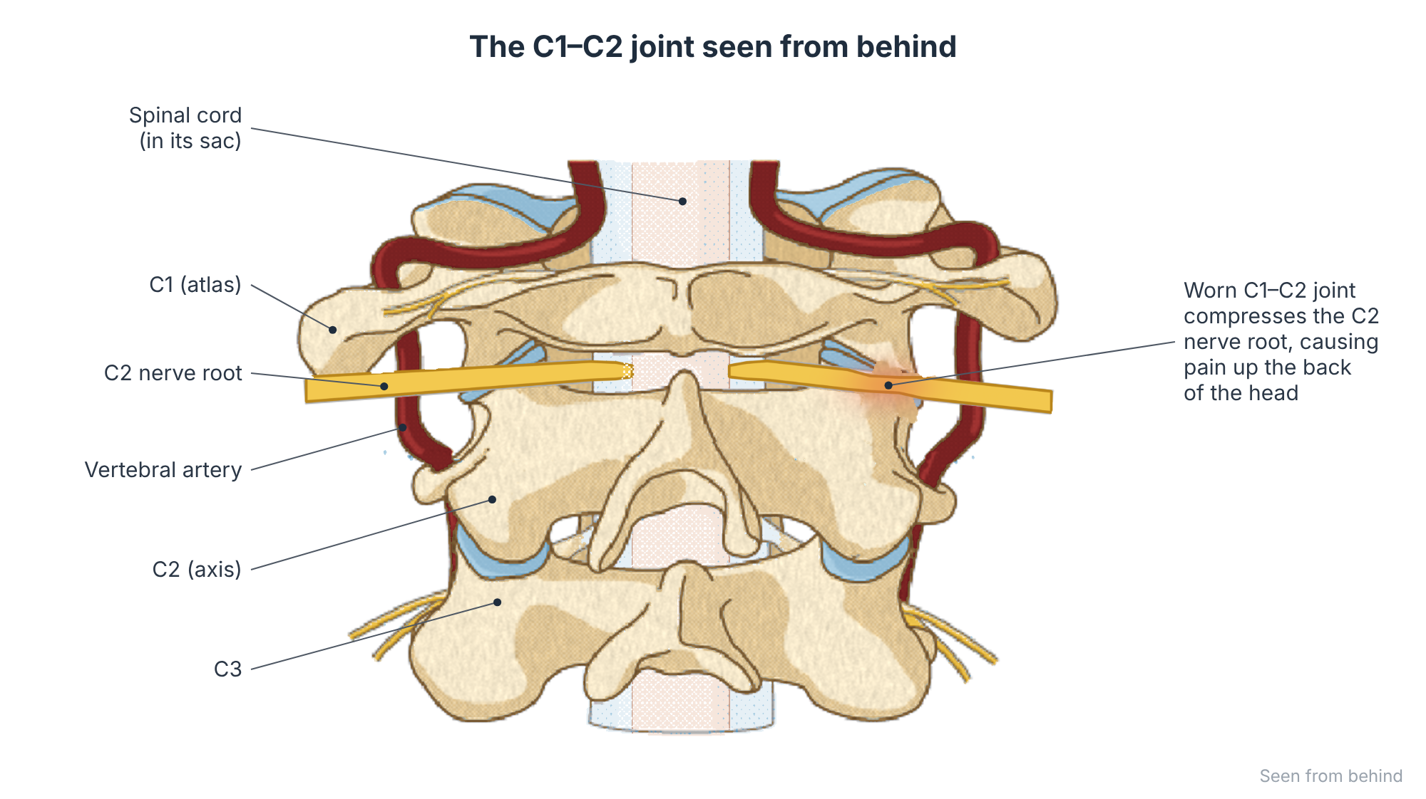
At a glance
- Most common in people over 60.
- Typically one-sided pain at the base of the skull, made much worse by turning the head.
- Often misdiagnosed as migraine or tension headache.
- Usually treated with medication, physiotherapy and targeted injections. Fusion of C1–C2 can be very effective for severe, persistent cases.
Symptoms
- Pain at the top of the neck and base of the skull, usually on one side.
- Pain spreading up the back of the head, sometimes towards the ear or behind the eye.
- Sharp, shooting or electric pains in the scalp (occipital neuralgia), and sometimes tenderness of the scalp.
- Marked pain and restriction when turning the head to one side, for example when reversing a car.
- Night pain in some patients.

Risk factors and causes
- Age-related osteoarthritis of the atlanto-axial joint.
- Inflammatory arthritis such as rheumatoid arthritis.
- Previous trauma to the upper neck.
- Occipital neuralgia can also arise from muscle tension, nerve entrapment, or rarely tumours or vascular problems, which is why careful assessment is needed.
How is it diagnosed?
Examination often shows a marked loss of rotation and tenderness over the C1–C2 joint or the occipital nerve. X-rays (including an open-mouth view), CT and sometimes SPECT-CT scans show arthritis and inflammation of the joint. A diagnostic injection of local anaesthetic into the C1–C2 joint or around the occipital nerve can confirm the pain source: if the pain disappears, the source is confirmed.
Non-surgical treatment
- Medication: anti-inflammatories and nerve-pain medicines such as amitriptyline, gabapentin or pregabalin.
- Physiotherapy and soft-tissue treatment for comfort. Avoid forcing rotation or stretching an arthritic C1–C2 joint: letting it settle and stiffen is often what eases the pain.
- Greater occipital nerve block: an injection of local anaesthetic and steroid near the nerve at the back of the head. It is quick and often gives weeks to months of relief.
- Image-guided C1–C2 joint injection with steroid.
- Pulsed radiofrequency treatment of the C2 nerve for recurrent pain.
Surgical options
Surgery is considered when pain is severe, clearly comes from the C1–C2 joint (confirmed by imaging and diagnostic injection), and has not responded to non-surgical treatment.
C1–C2 fusion (atlanto-axial arthrodesis)
The painful joint is fused using screws in C1 and C2 joined by rods (the Harms technique), or screws passed across the joint, usually through an incision at the back of the neck. Eliminating movement at the arthritic joint removes the source of pain.

What about other options?
Endoscopic techniques, disc replacement and lower-neck decompression do not apply here, because the C1–C2 joint has no disc and the problem is joint arthritis rather than nerve compression in the canal. Decompression of the occipital nerve itself is sometimes performed for pure nerve entrapment without joint arthritis.
What the evidence says: Results of C1–C2 fusion
The evidence comes mainly from case series rather than randomised trials, because the condition is relatively uncommon. In a long-term series (Ghanayem, Leventhal and Bohlman), the large majority of patients had substantial pain relief after C1–C2 fusion. The trade-off is a permanent loss of roughly half of the neck’s turning movement, which most patients adapt to well because their rotation was already painful and limited.
Recovery
After C1–C2 fusion, patients usually stay in hospital for 2 to 4 days and may wear a soft collar for comfort. Fusion takes about 3 months to become solid. Most people adapt to the reduced rotation by turning the shoulders and lower neck.
Talk to Prof Kruger
If you are living with this condition and would like an opinion on your options, you are welcome to book a consultation. Please bring any recent scans and reports.
References and key studies
- Ghanayem AJ, Leventhal M, Bohlman HH. Osteoarthrosis of the atlanto-axial joints: long-term follow-up after treatment with arthrodesis. J Bone Joint Surg Am. 1996;78(9):1300–1307.
- Harms J, Melcher RP. Posterior C1–C2 fusion with polyaxial screw and rod fixation. Spine. 2001;26(22):2467–2471.
- Headache Classification Committee of the International Headache Society. The International Classification of Headache Disorders, 3rd edition (ICHD-3): 13.4 Occipital neuralgia. Cephalalgia. 2018;38(1):1–211.
This page is general patient education based on published research and is not a substitute for a personal consultation. Treatment is always tailored to the individual.