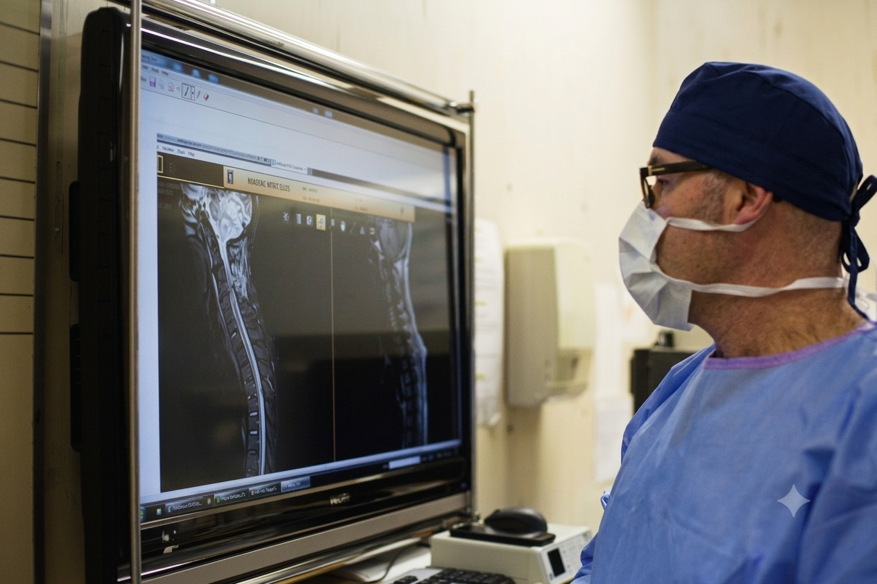
A Patient’s Guide to Spine Surgery and Procedures
Spinal Procedures – What to Expect
These guides explain common spinal procedures and what to expect before, during and after treatment. They are for general information only, and are not a substitute for a full discussion with Prof Kruger about your specific condition and surgery.
Spinal Injections
Nerve Root Block
A nerve root block is an injection into the sheath surrounding a nerve root, used both to reduce pain and to help pinpoint its source more precisely. A combination of steroid and local anaesthetic is used to settle pain and inflammation; relief can range from minimal to long-lasting, depending on your specific symptoms. You need to be experiencing symptoms at the time of the procedure for it to be effective – if your symptoms have settled beforehand, please let us know so the appointment can be rescheduled.
Before Your Procedure: Check with your doctor before stopping any medication. Bring previous imaging (MRI, CT, x-rays) if available. Arrange a driver, and let the nursing staff know if you are breastfeeding or there is a chance of pregnancy.
During the Procedure: You will remain awake throughout. A radiologist places anaesthetic and steroid around the nerve using x-ray (fluoroscopic) guidance, with contrast dye used to confirm needle position. Some pressure or discomfort is normal, and you may be asked how it compares with your usual pain.
After the Procedure: You may feel numbness or relief for up to 6 hours as the anaesthetic wears off; your usual symptoms may briefly return before the steroid takes effect over the next 2-3 days. If there is no change after a week, further investigation may be needed.
Possible side effects: facial flushing, mild fever, hiccups, insomnia, headache, fluid retention, increased appetite or heart rate, or abdominal cramping. These occur in around 5% of patients and usually settle within 1-3 days.
Caudal Epidural Injection
The epidural space surrounds the dura, the membrane covering the spinal cord and nerve roots. Nerves travel through this space down to the back and legs, and inflammation here, often from a damaged disc or contact with the bony spine, can cause pain in these areas. A caudal epidural places anti-inflammatory medicine into this space to settle nerve root inflammation, which may reduce pain in the back or legs while the underlying injury heals. It can provide lasting relief, or a period of several months of relief while things settle.
What Happens During the Procedure
An IV is started so relaxation medication can be given, and you will lie on your stomach on the x-ray table. After the skin is cleaned and numbed, a small needle is guided, using x-ray guidance, into the sacral hiatus, a small bony opening just above the buttock crease. Contrast dye confirms correct needle placement before a mixture of anaesthetic and anti-inflammatory (steroid) medicine is injected. These injections are usually given as a series of three, roughly two months apart.
Before and After
Nothing to eat or drink after 12pm the night before. Take your routine medications as usual (including blood pressure and diabetic medication), but let us know if you are on Coumadin or Glucophage, as timing may need to be adjusted. You will need a driver, as you will not be able to drive yourself home and your legs may feel weak or numb for a few hours. Afterwards you will be monitored in recovery for 30-60 minutes; most people return to normal activities, including work, the following day.
Discectomy
Discs degenerate naturally with age, and this can be influenced by genetics (a family history of back pain), smoking, and certain occupations. As a disc flattens and bulges, much like a worn car tyre, it can compress the nerve root behind it, or tear and leak disc material that irritates the nerve directly. Buttock and leg pain usually comes from this nerve compression or irritation, while axial back pain more often comes from the degenerate disc or facet joints themselves.
If symptoms persist for more than 6 weeks despite adequate conservative treatment, or if there are complications such as severe pain, bladder or bowel involvement, or progressive weakness, surgery is indicated.
A discectomy decompresses the prolapsed disc material from the neural canal. Only the prolapsed portion and any loose fragments are removed; the cavity then fills naturally with scar tissue. The operation addresses leg pain caused by nerve compression – it does not treat underlying back pain.
What to Expect
The operation itself takes about an hour. You will spend the first night in high-care and return to the ward the following day, where you are encouraged to mobilise out of bed. Most patients are discharged the next morning. Sutures are subcutaneous, though I like the knots snipped off a week later to avoid the occasional stitch abscess. Non-impact exercise can usually begin after 2 weeks, with a return to light duties and driving around the same time.
Possible Complications
| Complication | Details |
|---|---|
| Infection | 2-3% risk, minimised with sterile technique and preventive antibiotics. |
| Dural tear | 0.8-7.2% risk; may cause a post-operative headache and require a period of bed rest while the dura seals. |
| Nerve root injury | Usually related to nerve retraction during a difficult discectomy; generally settles with time. |
| Recurrent disc prolapse | Occurs in 5-15% of cases following successful surgery, a result of the underlying disc disease rather than the surgery itself. |
| Deep vein thrombosis (DVT) | A minimal risk overall, and rare in spinal surgery; minimised with TED stockings and anticoagulants where appropriate. |
| Residual numbness | Often remains after the pain of a pinched nerve resolves; this usually improves over time. |
| Epidural haematoma | Roughly 1% risk; a complication of post-operative bleeding that can compress bladder and bowel nerves and needs emergency evacuation. |
| Failure of surgery | Occasionally, nerve entrapment has become so chronic that established pain patterns do not fully resolve with decompression. |
| Ongoing lower back pain | May reflect ongoing degeneration of the spinal segment rather than a result of the surgery; occasionally a fusion is later needed to address the back pain component. |
Spinal fusion
You will be admitted through reception on the first floor once authorisation has taken place. Once on the ward, the nursing staff will prepare you for surgery with forms, documents and some skin preparation.
Depending on your co-morbidities, investigations may include ECG, cardiac Echo, blood tests, X-rays. A physician may also be involved in your co-management.
I will see you before your operation to confirm the surgery and indications, discuss any last minute concerns, and mark the more symptomatic side. Consent will be taken if this has not already been done, and the anaesthetist will usually also visit you at this time.
After surgery you will wake up in high-care/ICU and be fairly sedated, as is normal. Some discomfort is expected, but a combination of local anaesthetic in the wound and IV pain relief is usually adequate. I usually inject Fentanyl into your CSF space which gives excellent pain control for the first day. You will need to lie supine for 6 hours after surgery to allow the wound to seal and to allow clotting; thereafter you may sit up or turn onto your side as feels comfortable. A urinary catheter is normally inserted during surgery for convenience overnight, and depending on your age and any other conditions such as diabetes, a physician may help with your medical management.
Your Recovery Timeline
| Stage | What to Expect |
|---|---|
| Day 1-2 | Transferred back to the general ward and mobilized out of bed as you feel able, with a physiotherapist assisting twice daily. Normal diet resumes and the catheter is removed once you are comfortable standing. Patients having spinal injections usually go home the same day, and after smaller operations such as a discectomy most go home on day 2. |
| Day 3 | Most patients feel significantly better and the drip is removed. Physiotherapy continues twice daily. Post-op x-rays are taken and the wound dressing is checked; a water-proof dressing is applied for showering once dry. |
| Day 3-7 | Once your pain is controlled on oral medication, you are mobile, and you can manage stairs and the bathroom confidently, you may be discharged home. If needed, a step-down facility such as Palm Gardens can help you convalesce. During this time, if there is any leakage from your wound, fever, increasing discomfort, contact us or your GP immediately for advice. Short social walks are fine during this time. |
| Day 10 | Sutures are self-dissolving, but I prefer the knots snipped off to avoid the occasional stitch abscess – this can be done by yourself, your GP, or a pharmacy nurse. Dressings can usually be stopped after day 10. |
| Day 14 | The wound is healed. Showering can progress to bathing and swimming, and this is a good time to start gentle cardiovascular exercise such as spinning cycles. |
| Week 6 | X-rays are taken and reviewed. If any concerns I will see you for examination. Most patients are comfortable resuming normal walking and non-impact exercise. |
| Month 3 | Final x-rays are taken for uncomplicated fusions. If normal, no further imaging is needed and you may return to any activity you enjoy, including impact sports. |
Returning to Normal Life
Return to Work: Sedentary or office work can often resume on light duties after 2 weeks. I am happy to support an early return, or can book you off for 6 weeks if needed. Normal duties usually resume after 3 months for uncomplicated surgery.
Driving: You may drive if you have no neurological deficit, are not taking strong opioid painkillers, and can safely perform an emergency stop – often around 2 weeks, depending on comfort.
Sitting: You may sit in whichever position is comfortable for you; there is no restriction on sitting posture.
Flying: Short (2 hour) flights are usually fine after discharge if there is no family history of DVT. For international flights, 4 weeks post-surgery is generally allowed, though this can be discussed individually.
Long-Term Advice
In the long term, I do not believe specific “core” exercises are necessary, and they may even aggravate the levels around your fusion – there is no strong scientific evidence supporting them. I recommend sticking to normal exercise you enjoy, and avoiding sit-ups, planking, or back-loading movements.
Because of the fusion, there is a modestly increased chance (roughly 20% over several years) that another level may eventually need attention. This is usually a reflection of your spine’s general condition rather than a consequence of the surgery itself, similar to how one filling does not cause the next cavity, but general tooth decay does.
Smoking measurably increases the risk of infection, non-fusion, poor wound healing, and worse long-term spinal health, so quitting, or at least cutting down, around the time of surgery is strongly advised.
Exercise Recommendations
| Recommended | Best Avoided |
|---|---|
| Non-impact cardio (swimming, stationary cycling) | Core-specific exercises, planking, sit-ups |
| Exercise you enjoy and can sustain | Stretching or spinal loading drills |
| Listening to your body and your back | Ignoring persistent or worsening pain |
When to Contact Us
If you develop a fever, wound discharge, or increasing back or leg pain that persists at any stage of your recovery, please contact the rooms – this may need blood tests, antibiotics, or further imaging. If you have metal instrumentation in your spine, a proven wound infection is usually treated with 6 weeks of antibiotics.
Spinal Health and Common Sense
A Personal Perspective…
Spinal pain is part of the human condition – most of us will experience it at some point, and thankfully around 90% of episodes settle within 6-8 weeks, regardless of treatment. It is a natural consequence of an ageing spine: joint spaces narrow, discs lose hydration and soften, and osteophytes form as the spinal column stiffens with age. Interestingly, spinal pain tends to peak in our middle decades and then ease again after our 50s and 60s, even as degeneration on scans continues to progress.
Very few remedies for ordinary spinal pain are proven to outperform this natural history. Physiotherapy, chiropractic care, massage and similar treatments do not necessarily resolve pain faster than time alone, but they do offer valuable reassurance and support while symptoms settle.
Where my view differs from the mainstream is around exercise and “core stability”. There is a common assumption that spinal pain results from weak core muscles or poor posture, and that strengthening and stretching will fix a degenerating spine. I do not think the evidence supports this – much like an arthritic knee is not treated by strengthening the muscles around it, I do not believe a degenerating spinal joint benefits from targeted core work or stretching. Elite athletes and fitness professionals with excellent strength and conditioning still develop spinal degeneration and pain, which supports this view.
My advice, which applies mainly to patients who have not improved despite standard treatment, is to let a degenerating spine follow its natural course toward stiffening, rather than fighting it with core exercises, stretching, or spinal loading drills. I am a strong advocate for exercise, but favour non-impact cardiovascular activity such as swimming or stationary cycling, which lifts mood and supports overall health without loading the spine unnecessarily. Maintaining a healthy weight also plays an important, often underestimated, role in managing back pain.
Our spines are very closely linked to systemic health, and with cardiovascular disease, diabetes and autoimmune diseases, our spines similarly age rapidly. Biological clocks are nowadays accurately measured by proteomic clocks, and these are accelerated with ill health, obesity and smoking. Healthy weight and regular cardiovascular exercise slow down your biological ageing, and prolong your spine longevity.