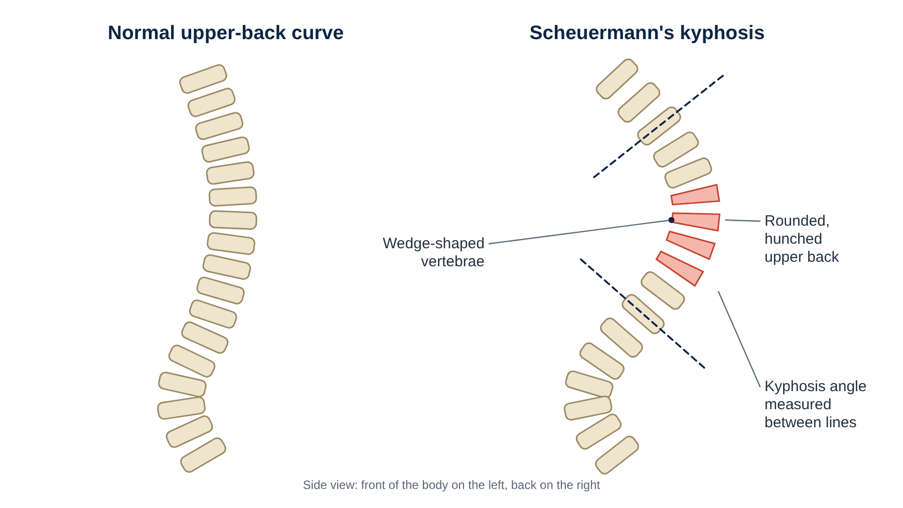
Scheuermann’s kyphosis is a condition in which several vertebrae in the upper back grow into a wedge shape during adolescence. Together, these wedges create a rounded, hunched upper back that cannot be corrected simply by standing up straight.
At a glance
- Usually noticed between the ages of 10 and 16, often as “poor posture” that does not correct.
- Defined by at least three neighbouring vertebrae each wedged by 5 degrees or more.
- Most people need only exercise and monitoring. Bracing helps during growth.
- Surgery is considered for severe curves (typically more than 70 to 75 degrees), persistent pain or neurological problems.
What causes it?
The exact cause is unknown. There is a strong genetic component, and it is thought that the growth plates at the front of the vertebrae grow more slowly than those at the back, creating wedge-shaped bones. Small disc herniations into the vertebrae (Schmorl’s nodes) and irregular endplates are typical on X-ray.
Symptoms
- A rounded or humped upper back that stays curved even when lying flat or trying to stand straight.
- Aching pain between the shoulder blades, worse with activity or prolonged sitting.
- Tight hamstrings and chest muscles.
- An increased compensatory curve in the lower back, which can cause low back pain in adulthood.
- Very rarely, pressure on the spinal cord, causing leg weakness.
Risk factors
- Family history.
- Being male (it is about twice as common in boys).
- Adolescent growth spurt.
- Possibly heavy loading during growth.
How is it diagnosed?
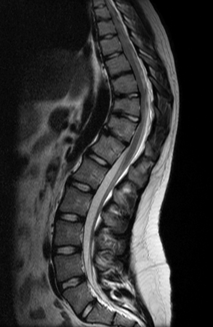
A clinical examination includes the forward-bending test, where the rigid curve becomes obvious. A standing side-view X-ray of the whole spine measures the kyphosis angle and shows the wedged vertebrae. MRI is used if there are neurological symptoms or before surgery.
Non-surgical treatment

- Exercise: regular non-impact cardio such as cycling and swimming. Stretching and core programmes do not correct the wedge-shaped vertebrae and are not needed.
- Bracing: in growing adolescents with significant curves (often 60 to 75 degrees), a brace worn for many hours a day can partially correct the wedging while the spine is still growing.
- Pain management with simple analgesics and activity modification.
- Monitoring with periodic X-rays until growth is complete.
Surgical options
Surgery is considered for curves larger than about 70 to 75 degrees, persistent pain despite treatment, progressive deformity, neurological problems or significant concerns about appearance.
Posterior spinal fusion with osteotomies
The standard operation is performed through the back. Small wedges of bone are removed at several levels (Ponte or Smith-Petersen osteotomies) to make the spine flexible. Pedicle screws and rods are then used to gradually straighten the curve, and the spine is fused in the corrected position. Spinal cord monitoring is used throughout the operation.
What about other techniques?
Endoscopic surgery, isolated decompression and disc replacement do not correct this deformity and are not used. Combined front-and-back surgery, once common, is now rarely required thanks to modern pedicle screw systems.
What the evidence says: Natural history
A long-term follow-up study of untreated patients (Murray, Weinstein and Spratt, JBJS 1993) found that adults with Scheuermann’s kyphosis had more back pain and more physically limited jobs than average, but most functioned well and had little interference with daily activities. This supports a conservative approach for most patients.
What the evidence says: Bracing
In a series of patients treated with a Milwaukee brace during growth (Sachs and colleagues, JBJS 1987), most showed improvement or stabilisation of their kyphosis, although curves greater than 74 degrees were more likely to progress or need surgery.
Recovery after surgery
Hospital stay is usually 4 to 6 days. School or desk work can typically resume after 4 to 6 weeks, with contact sports deferred until the fusion is solid, usually at 6 to 12 months. Most patients report better posture, pain relief and high satisfaction.
Talk to Prof Kruger
If you are living with this condition and would like an opinion on your options, you are welcome to book a consultation. Please bring any recent scans and reports.
References and key studies
- Sørensen KH. Scheuermann’s juvenile kyphosis: clinical appearances, radiography, aetiology, and prognosis. Copenhagen: Munksgaard; 1964.
- Murray PM, Weinstein SL, Spratt KF. The natural history and long-term follow-up of Scheuermann kyphosis. J Bone Joint Surg Am. 1993;75(2):236–248.
- Sachs B, et al. Scheuermann kyphosis: follow-up of Milwaukee-brace treatment. J Bone Joint Surg Am. 1987;69(1):50–57.
- Lowe TG, Line BG. Evidence based medicine: analysis of Scheuermann kyphosis. Spine. 2007;32(19 Suppl):S115–S119.
This page is general patient education based on published research and is not a substitute for a personal consultation. Treatment is always tailored to the individual.