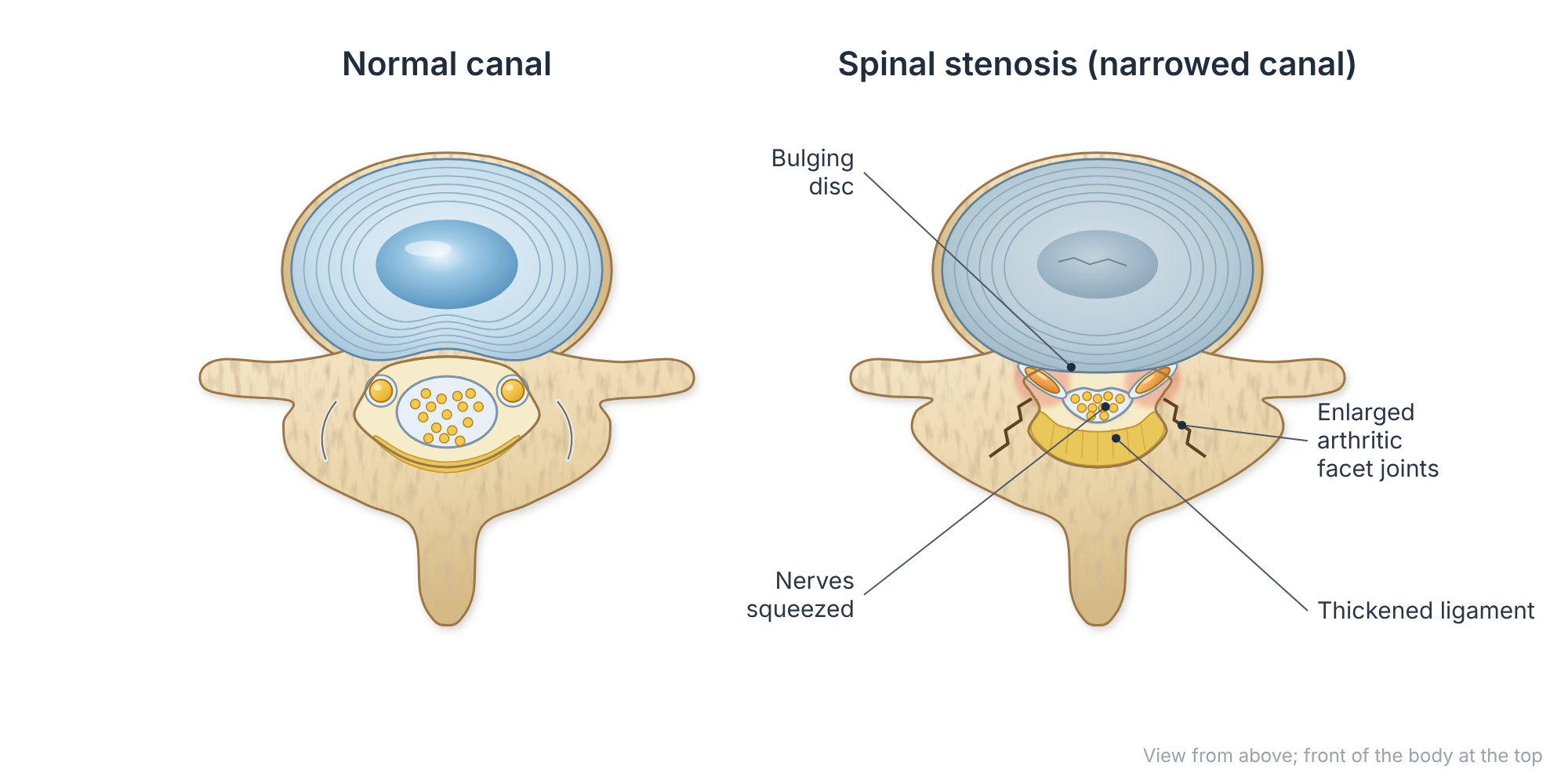
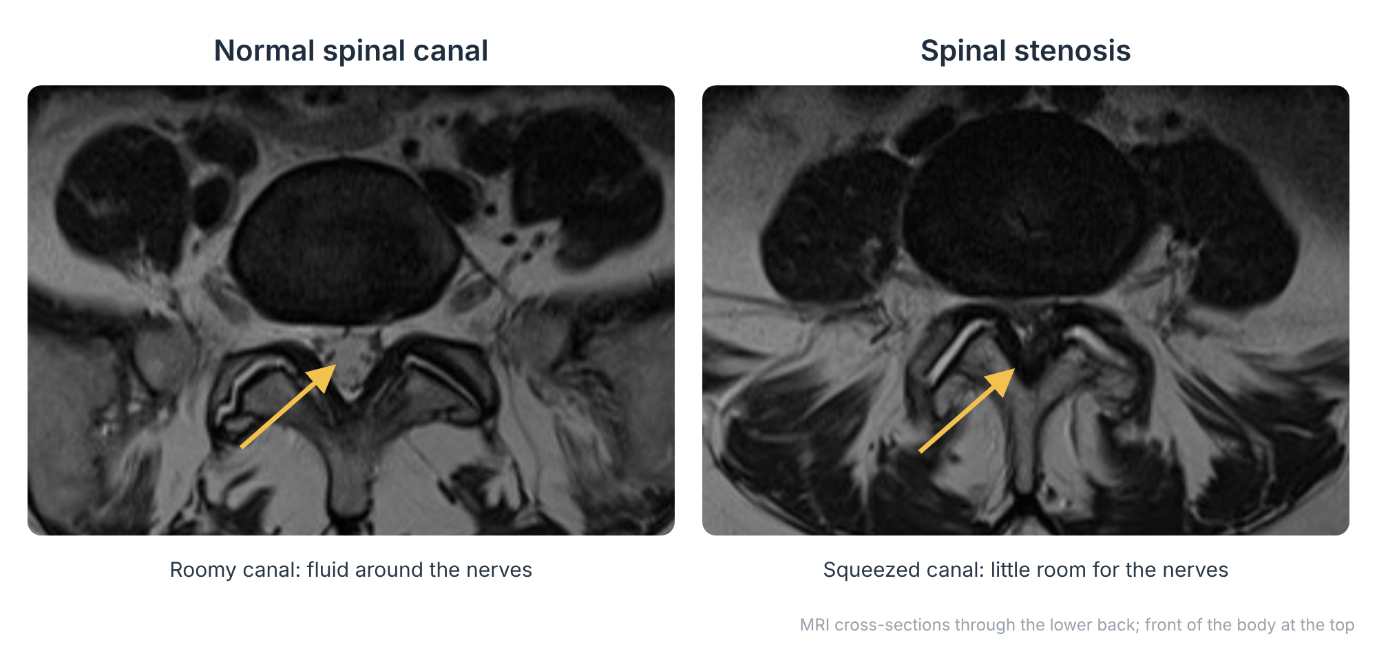
Lumbar spinal stenosis means the tunnel that carries the nerves through the lower back has become narrowed. It develops slowly with age as discs bulge, the small joints enlarge with arthritis and ligaments thicken. The nerves are squeezed, especially when standing and walking.
At a glance
- The most common reason for spinal surgery in people over 65.
- Classic symptom: leg pain, heaviness or numbness that comes on with walking or standing and eases with sitting or bending forward.
- Many people stay stable for years and manage well without surgery.
- Decompression surgery reliably improves walking distance and leg pain when symptoms limit daily life.
Symptoms
- Neurogenic claudication: pain, cramping, tingling or heaviness in the buttocks and legs after walking a certain distance or standing for a while.
- Relief from sitting down or leaning forward, for example on a shopping trolley (the “shopping-trolley sign”). Cycling is often easier than walking.
- Numbness or weakness in the legs.
- Lower back ache and stiffness.
- Rarely, problems with bladder or bowel control. This needs urgent assessment.
Stenosis can be confused with poor circulation to the legs (vascular claudication). With circulation problems, standing still is enough to relieve the pain, and foot pulses are often weak. Your doctor may check both.
Risk factors
- Age over 50.
- Degenerative arthritis of the spine and disc degeneration.
- Spondylolisthesis (a slipped vertebra).
- A naturally narrow canal (congenital stenosis), which brings symptoms on at a younger age.
- Previous spinal surgery or injury, and adult scoliosis.
How is it diagnosed?
Diagnosis is based on your symptoms, an examination and an MRI scan, which shows the degree and level of narrowing. Standing X-rays, including flexion and extension views, check for slip or instability, which can influence the choice of surgery.
Non-surgical treatment
- Non-impact cardio: stationary cycling (often comfortable because you lean forward) and swimming. Physiotherapy can help with comfort, but stretching and core programmes are not needed.
- Medication: simple analgesics and anti-inflammatories. Nerve-pain medication helps some patients.
- Epidural steroid injections: may give temporary relief. A large US trial found little added benefit over local anaesthetic alone, so they are used selectively.
- Weight control and staying active, which help to maintain walking tolerance.
Surgical options
Surgery is considered when symptoms significantly limit walking or daily activities despite conservative treatment, or if there is progressive weakness. The aim is to make more room for the nerves (decompression).

Endoscopic and minimally invasive decompression
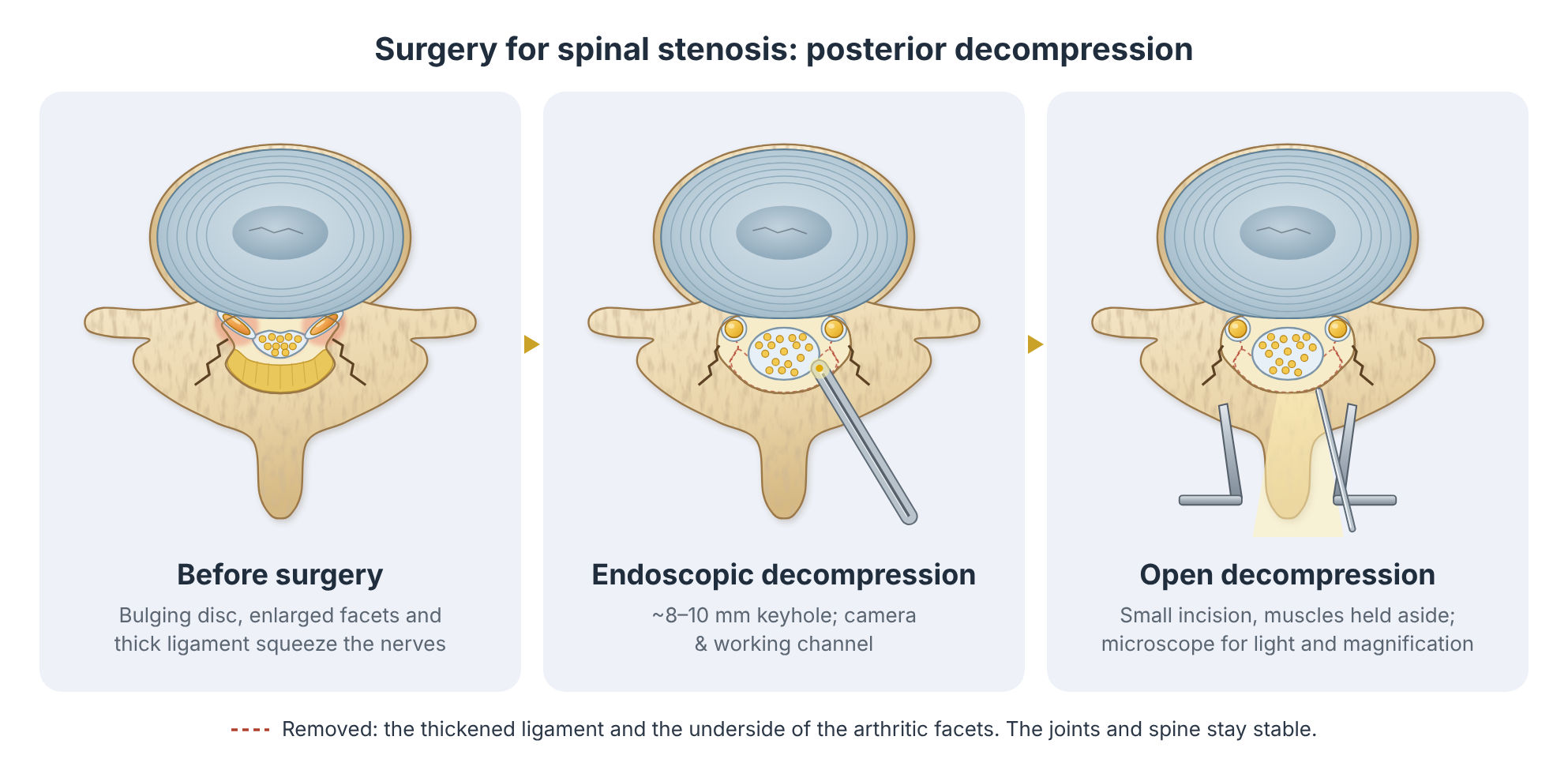
Using an endoscope or tubular retractors, the surgeon removes thickened ligament and bone from one side and “undercuts” across to decompress both sides. This preserves the midline structures and muscle, with less pain and a quicker return home.
Open microsurgical decompression (laminectomy or laminotomy)
The traditional and very effective operation. Bone and ligament pressing on the nerves are removed under the microscope, while preserving the joints that give the spine stability.
Decompression with fusion
Fusion is added only when the spine is unstable, there is a significant slip that moves, or there is a deformity such as scoliosis. For stenosis without instability, fusion adds cost and risk without adding benefit.
Disc replacement
Disc replacement is not used to treat spinal stenosis, which is mainly caused by arthritis of the facet joints and thickened ligament.
What the evidence says: The SPORT stenosis trial
In SPORT, patients with spinal stenosis treated with decompression surgery had significantly greater improvement in pain and function than those treated non-surgically, and the advantage persisted at 4 and 8 years. In a separate trial (Delitto and colleagues, 2015), surgery and a structured physiotherapy programme gave similar results at 2 years, although many physiotherapy patients later chose surgery.
What the evidence says: Is fusion needed?
The Swedish Spinal Stenosis Study (Försth and colleagues, NEJM 2016) randomised 247 patients to decompression alone or decompression plus fusion. Adding fusion did not improve results at 2 or 5 years, even in patients with a mild degenerative slip, but it made surgery longer and more costly. Decompression alone is therefore the standard for most patients with stenosis.
Recovery
Most people walk on the day of surgery and go home within 1 to 2 days. Leg symptoms often improve quickly, while numbness can take months to recover. Around 7 to 8 out of 10 patients report a good result, and walking distance usually improves markedly. Some people need further surgery in later years as degeneration continues at other levels.
Talk to Prof Kruger
If you are living with this condition and would like an opinion on your options, you are welcome to book a consultation. Please bring any recent scans and reports.
References and key studies
- Weinstein JN, et al. Surgical versus nonsurgical therapy for lumbar spinal stenosis. N Engl J Med. 2008;358(8):794–810.
- Weinstein JN, et al. Surgical versus nonoperative treatment for lumbar spinal stenosis four-year results of the Spine Patient Outcomes Research Trial. Spine. 2010;35(14):1329–1338.
- Lurie JD, et al. Long-term outcomes of lumbar spinal stenosis: eight-year results of the Spine Patient Outcomes Research Trial (SPORT). Spine. 2015;40(2):63–76.
- Försth P, et al. A randomized, controlled trial of fusion surgery for lumbar spinal stenosis. N Engl J Med. 2016;374(15):1413–1423.
- Delitto A, et al. Surgery versus nonsurgical treatment of lumbar spinal stenosis: a randomized trial. Ann Intern Med. 2015;162(7):465–473.
- Friedly JL, et al. A randomized trial of epidural glucocorticoid injections for spinal stenosis. N Engl J Med. 2014;371(1):11–21.
This page is general patient education based on published research and is not a substitute for a personal consultation. Treatment is always tailored to the individual.