The thoracolumbar junction, where the stiff mid-back meets the more mobile lower back (around T11 to L2), is the most common site of spinal fractures. In Cape Town, these injuries are commonly seen after kiteboarding, paragliding, mountain-biking and horse-riding accidents, where a hard landing on the feet or buttocks drives a heavy load straight down the spine.

At a glance
- Wedge (compression) fractures: the front of the vertebra is squashed but the back wall is intact. They are stable and usually heal without surgery.
- Burst fractures: the entire vertebral body breaks under a heavy vertical load, and fragments may be pushed back into the spinal canal.
- Many burst fractures without nerve injury can also be treated without surgery.
- Surgery is needed for fractures with nerve or spinal cord injury, damage to the ligaments at the back of the spine, or significant instability or deformity.
How these injuries happen
In kiteboarding, a “kite-loop” or a gust can lift a rider several metres before an uncontrolled landing. In paragliding, collapse of the wing close to the ground or a heavy landing during take-off or landing transmits force straight up the spine. Similar forces occur in falls from height, road accidents and horse riding. The spine fails at the thoracolumbar junction because this is where a rigid segment meets a flexible one.
Symptoms
- Immediate, severe back pain after the injury.
- Pain on standing, walking or moving, sometimes with a visible step or bump in the back.
- Bruising over the spine.
- Numbness, tingling or weakness in the legs, or difficulty passing urine. These suggest nerve or spinal cord injury and are an emergency.
After a high-energy fall
Anyone with back pain after a significant fall, especially with leg numbness or weakness, should be kept still and assessed at a hospital emergency unit. A careful neurological examination and CT scan are essential.
Risk factors
- Extreme and adventure sports: kiteboarding, paragliding, skydiving, motocross and mountain-biking.
- Falls from height and motor vehicle accidents.
- Osteoporosis, which allows fractures from much smaller forces (see osteoporotic spinal fractures).
- Conditions that stiffen the spine, such as ankylosing spondylitis, which make fractures more unstable.
How is it diagnosed?
A CT scan shows the bony injury in detail and how much the canal is narrowed. MRI shows injury to the spinal cord, nerves and the ligaments at the back of the spine (the posterior ligamentous complex), which are critical for stability. Fractures are graded using the AO Spine and TLICS classification systems, which combine the fracture pattern, the neurological status and the integrity of the ligaments to guide treatment.
Non-surgical treatment
- Wedge fractures: pain relief, early mobilisation and physiotherapy. Most heal within 8 to 12 weeks.
- Stable burst fractures without nerve injury: early walking with pain control, with or without a brace, and regular X-rays to make sure the alignment holds.
- Physiotherapy to restore fitness and return to sport, usually after 3 to 6 months once healing is confirmed.
What the evidence says: Do stable burst fractures need surgery?
In a randomised trial of burst fractures without neurological injury (Wood and colleagues, JBJS 2003), non-surgical treatment gave pain and function results equal to or better than surgical fusion, with fewer complications. Long-term follow-up at 16 to 22 years confirmed similar or better outcomes with non-surgical care. A Canadian trial (Bailey and colleagues, Spine Journal 2014) found that a brace was not necessary for these fractures: patients who did not wear one did just as well.
Surgical options
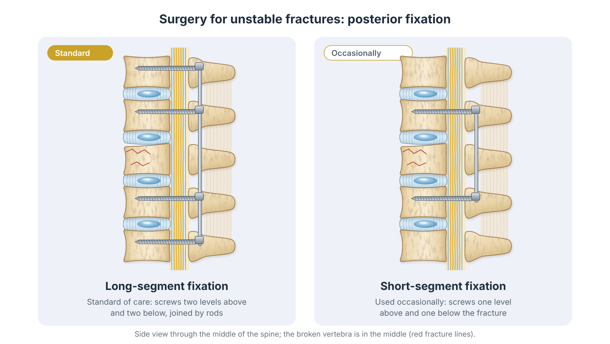
Surgery is recommended for unstable fractures, fractures with neurological injury, injury to the posterior ligaments, severe kyphotic deformity, or when early mobilisation is important (for example, with multiple injuries).

Minimally invasive (percutaneous) fixation
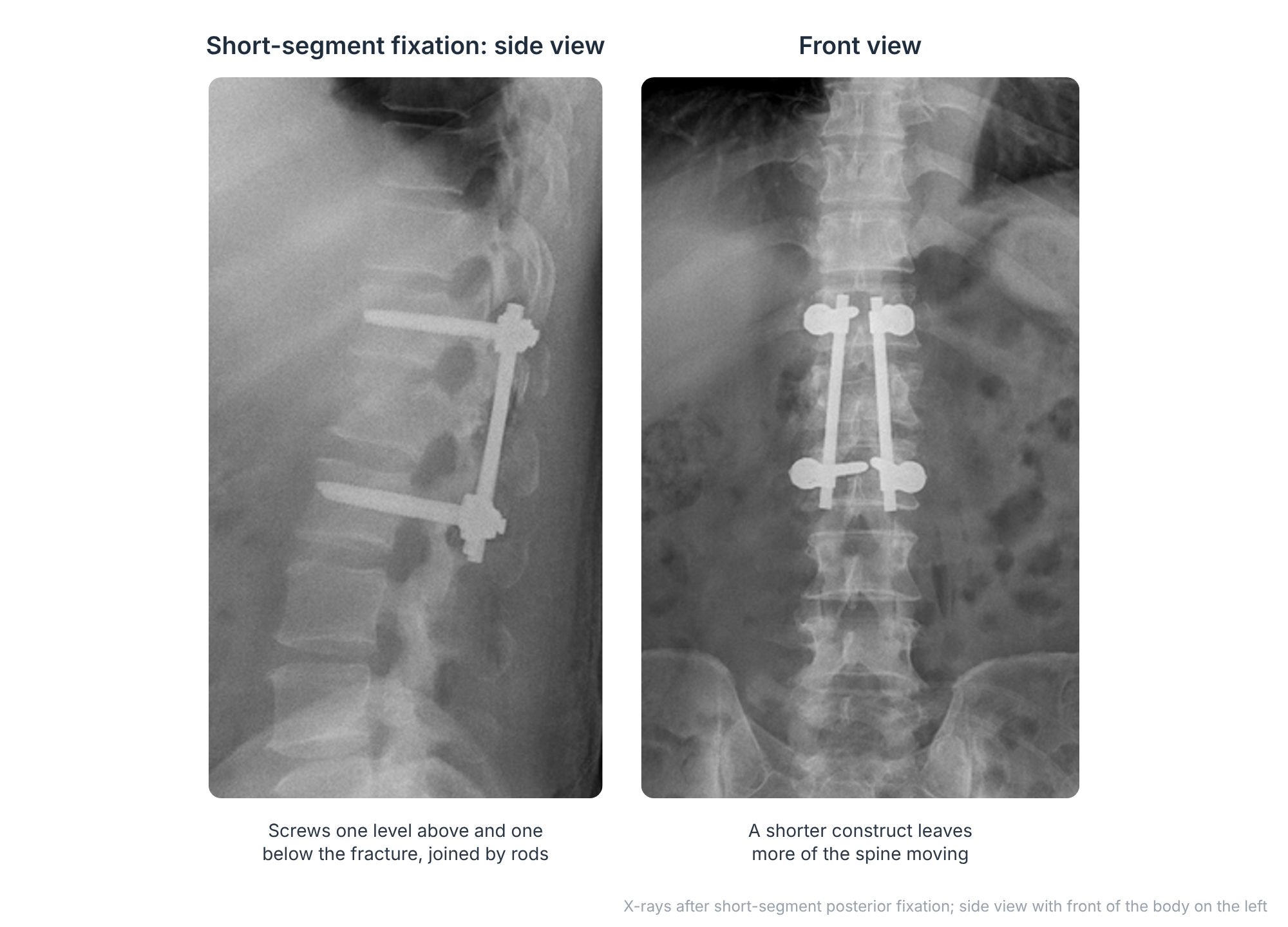
Screws are inserted through small incisions into the vertebrae above and below the fracture and joined with rods passed under the skin. This stabilises the spine with less muscle damage, less blood loss and faster recovery. In young patients, the implants can sometimes be removed after the fracture has healed, restoring movement.
Open decompression and fusion
When fragments of bone are pressing on the spinal cord or nerves, the canal is opened and the fragments are removed or pushed back (decompression), followed by fixation with screws and rods. In some cases, the broken vertebral body is replaced with a cage through an approach from the side or front.
Spinal cord injury
Patients with spinal cord injury are treated as an emergency in a specialised spinal cord injury unit. Evidence suggests that early decompression, within 24 hours where possible, improves the chance of neurological recovery.
What the evidence says: Timing in spinal cord injury
The STASCIS study (Fehlings and colleagues, PLoS One 2012) found that patients with spinal cord injury in the neck who had decompression within 24 hours were more likely to achieve a meaningful improvement in neurological function than those decompressed later. Similar principles of early decompression are applied to injuries of the thoracolumbar spine.
Recovery and returning to sport
Most patients with stable fractures return to normal daily activities within 6 to 12 weeks. A return to high-risk sports such as kiteboarding or paragliding is usually possible after 4 to 6 months, once healing is confirmed, fitness is restored and the patient is pain-free. After fusion, the timing depends on the extent of surgery.
Complex fractures
High-energy injuries can cause complex fractures that need specialised reconstruction. In this example, a fall from height broke the sacrum in several places, separating the lower spine from the pelvis (lumbo-sacral dissociation). It was treated with lumbo-sacral fixation, joining the lower lumbar spine to the pelvis with screws and rods.

Talk to Prof Kruger
If you are living with this condition and would like an opinion on your options, you are welcome to book a consultation. Please bring any recent scans and reports.
References and key studies
- Vaccaro AR, et al. A new classification of thoracolumbar injuries: the importance of injury morphology, the integrity of the posterior ligamentous complex, and neurologic status (TLICS). Spine. 2005;30(20):2325–2333.
- Vaccaro AR, et al. AOSpine thoracolumbar spine injury classification system. Spine. 2013;38(23):2028–2037.
- Wood K, et al. Operative compared with nonoperative treatment of a thoracolumbar burst fracture without neurological deficit: a prospective, randomized study. J Bone Joint Surg Am. 2003;85(5):773–781.
- Wood KB, et al. Operative compared with nonoperative treatment of a thoracolumbar burst fracture without neurological deficit: a prospective randomized study with follow-up at sixteen to twenty-two years. J Bone Joint Surg Am. 2015;97(1):3–9.
- Bailey CS, et al. Orthosis versus no orthosis for the treatment of thoracolumbar burst fractures without neurologic injury: a multicenter prospective randomized equivalence trial. Spine J. 2014;14(11):2557–2564.
- Fehlings MG, et al. Early versus delayed decompression for traumatic cervical spinal cord injury: results of the Surgical Timing in Acute Spinal Cord Injury Study (STASCIS). PLoS One. 2012;7(2):e32037.
This page is general patient education based on published research and is not a substitute for a personal consultation. Treatment is always tailored to the individual.