Spinal metastases are cancer deposits that have spread to the spine from elsewhere in the body. The spine is the most common site of bone spread. Metastases can cause pain, weaken the vertebrae so that they fracture, and, if tumour grows into the spinal canal, compress the spinal cord. Prompt recognition and treatment can preserve the ability to walk and greatly improve quality of life.

At a glance
- Up to 30 to 40% of people with cancer develop spinal metastases.
- The most common primary cancers are breast, prostate, lung, kidney and thyroid.
- New back pain in anyone with a history of cancer should be investigated.
- Metastatic spinal cord compression is an emergency: treatment within hours can make the difference between walking and paralysis.
Symptoms
- New back or neck pain that is constant, worse at night or when lying flat, and does not settle with rest.
- Band-like pain around the chest or abdomen.
- Sudden pain from a fracture.
- Numbness, heaviness or weakness of the legs, unsteadiness, or difficulty with bladder or bowel control. These suggest spinal cord compression.
Metastatic spinal cord compression is an emergency
If you have cancer and develop new leg weakness, numbness, walking difficulty or bladder or bowel problems, seek emergency care immediately. Steroids (dexamethasone) and urgent MRI are started straight away, and treatment ideally begins within 24 hours.
Risk factors
- A current or previous diagnosis of cancer, especially breast, prostate, lung, kidney or thyroid cancer.
- Advanced-stage cancer or known spread to other bones.
- Sometimes, a spinal metastasis is the first sign of an undiagnosed cancer.
How is it diagnosed?
MRI of the whole spine shows all deposits and any cord compression. CT assesses bone destruction and stability, and PET-CT or bone scans stage the disease. A biopsy is performed if the primary cancer is unknown.
Treatment: a team decision
Treatment is planned by a multidisciplinary team of oncologists, radiation oncologists and spinal surgeons. Decisions consider four key questions (the NOMS framework): the Neurological status, the Oncological behaviour of the tumour (especially sensitivity to radiotherapy), the Mechanical stability of the spine, and the Systemic health of the patient. Instability is scored using SINS, and prognosis using tools such as the revised Tokuhashi score.
Non-surgical treatment
- Radiotherapy: conventional radiotherapy for pain and radiosensitive tumours, and stereotactic body radiotherapy (SBRT), which delivers high, precise doses that can control even “radioresistant” tumours such as kidney cancer.
- Systemic treatment: chemotherapy, hormone therapy, targeted therapy and immunotherapy, which control disease throughout the body.
- Bone-protective drugs: zoledronic acid or denosumab reduce fractures and pain.
- Pain management and bracing for comfort.
Cement augmentation
Kyphoplasty or vertebroplasty can relieve pain from a fracture caused by metastases, often within days, with minimal recovery time (see the CAFE trial on the multiple myeloma page). It is often combined with radiotherapy.
Surgery
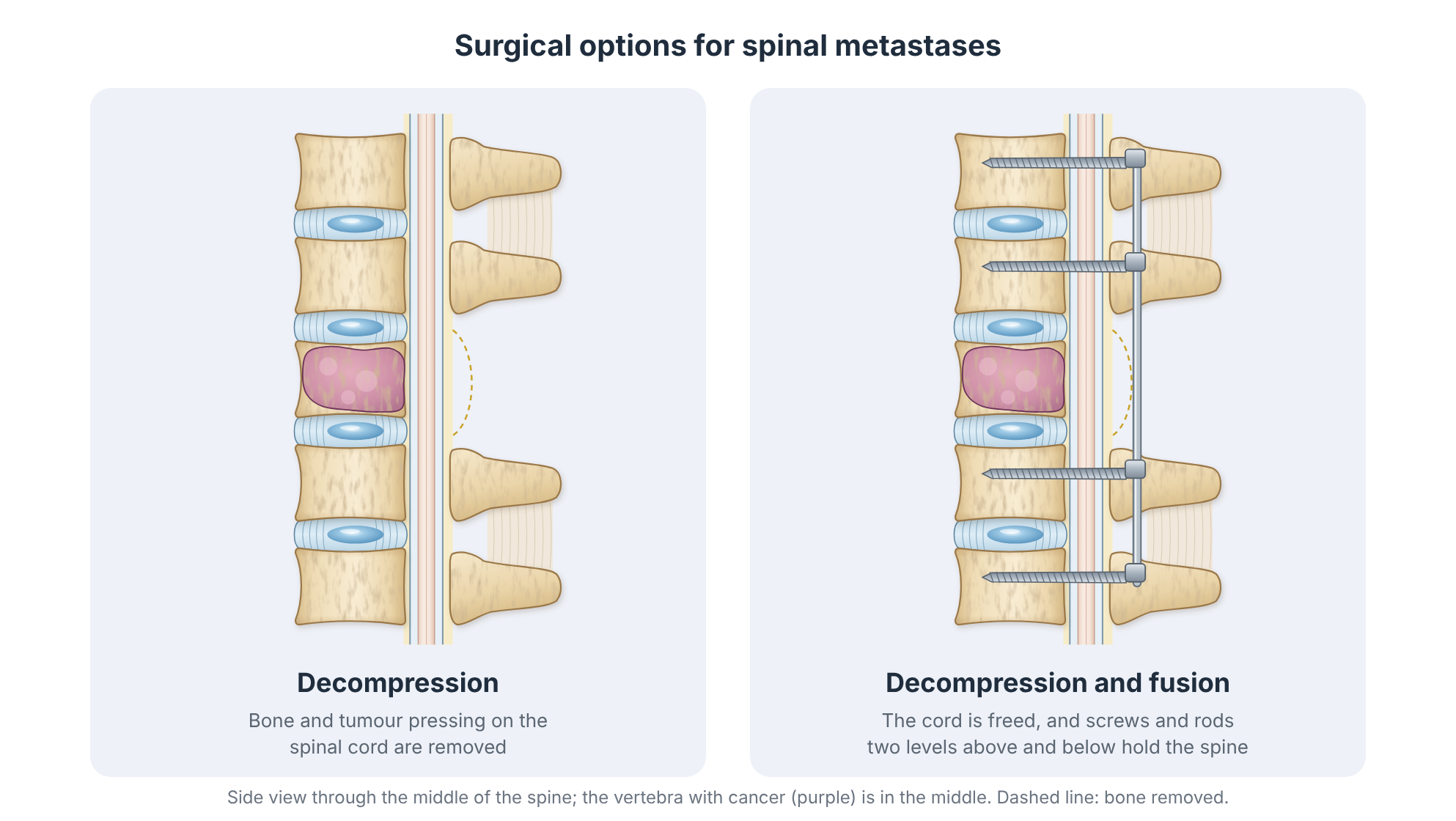
Surgery is considered for spinal instability, spinal cord compression (especially from radioresistant tumours or bone fragments), or progressive neurological deficit, in patients well enough to benefit.
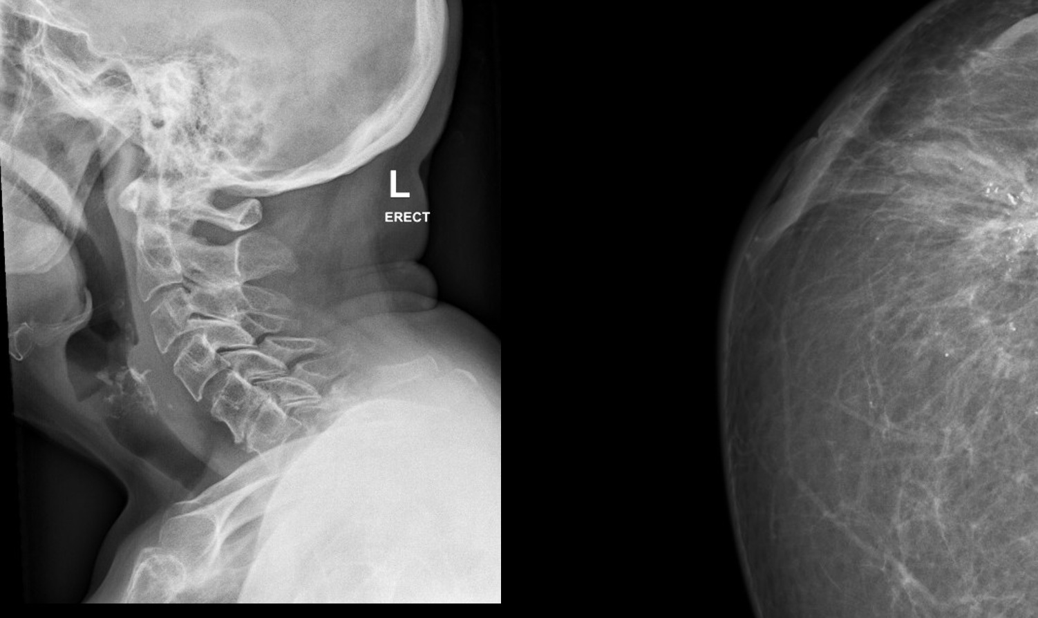
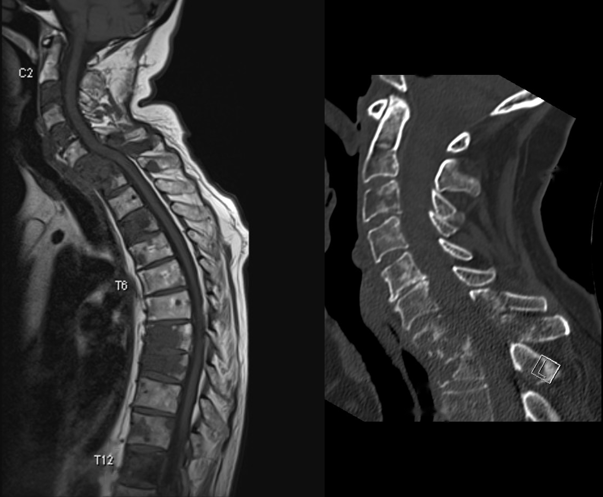
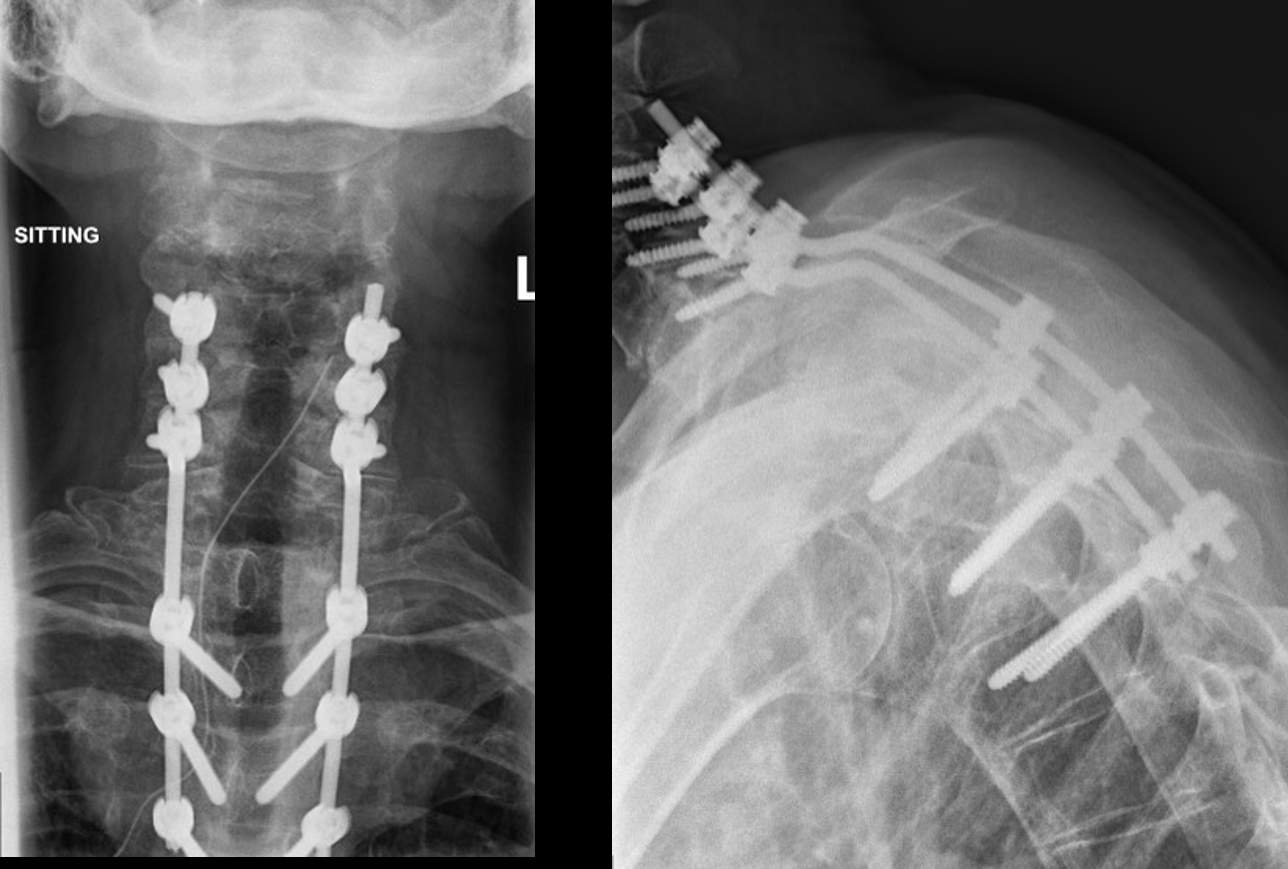
Example: breast cancer spread to the spine
Previous breast cancer is unfortunately a common source of spread to the spine. In this patient the MRI and CT showed the extent of bone destruction, with a threat of paralysis. Spinal instrumentation (screws and rods) helps to keep the patient walking and prevents the spine from progressively collapsing forward (kyphosis) and compressing the spinal cord.
Example: not all spinal tumours are cancer
Not all spinal tumours are malignant. This young girl had a benign spinal tumour that caused paralysis of her legs (paraplegia) and complete loss of bladder and bowel control. The entire tumour was removed and the spine was reconstructed with screws and rods from behind and a cage in front. The cage is made of PEEK, a strong plastic that does not show up clearly on X-rays. She made a full neurological recovery.


Show surgical photos (graphic)

- Minimally invasive (percutaneous) stabilisation: screws and rods inserted through small incisions, often with cement reinforcement, to stabilise the spine with rapid recovery so that radiotherapy or systemic treatment can start quickly.
- “Separation surgery”: the tumour is carefully removed from around the spinal cord and the spine is stabilised, creating a safe margin for high-dose SBRT to treat the remaining tumour.
- Open decompression and fusion, or vertebral body reconstruction: for more extensive tumours. Occasionally, complete removal of a solitary metastasis is performed (en-bloc resection) in selected cases such as isolated kidney or thyroid cancer metastases.
What the evidence says: Surgery plus radiotherapy
In a landmark randomised trial (Patchell and colleagues, Lancet 2005) of patients with metastatic spinal cord compression, those treated with decompressive surgery followed by radiotherapy were significantly more likely to walk after treatment (84% versus 57%) and to keep walking for longer than those treated with radiotherapy alone. Patients who could not walk at the start were also more likely to regain walking with surgery.
What the evidence says: Stereotactic radiotherapy
The SC.24 trial (Sahgal and colleagues, Lancet Oncology 2021) found that stereotactic radiotherapy gave better complete pain relief than conventional radiotherapy for painful spinal metastases at 3 and 6 months.
Outlook
The outlook depends mainly on the type of cancer and how well it responds to treatment. For many people, especially those with breast, prostate, kidney or thyroid cancer, modern treatment allows years of good-quality life. Preserving the ability to walk and controlling pain are central goals of spinal treatment.
Talk to Prof Kruger
If you are living with this condition and would like an opinion on your options, you are welcome to book a consultation. Please bring any recent scans and reports.
References and key studies
- Patchell RA, et al. Direct decompressive surgical resection in the treatment of spinal cord compression caused by metastatic cancer: a randomised trial. Lancet. 2005;366(9486):643–648.
- Laufer I, et al. The NOMS framework: approach to the treatment of spinal metastatic tumors. Oncologist. 2013;18(6):744–751.
- Fisher CG, et al. A novel classification system for spinal instability in neoplastic disease (SINS). Spine. 2010;35(22):E1221–E1229.
- Tokuhashi Y, et al. A revised scoring system for preoperative evaluation of metastatic spine tumor prognosis. Spine. 2005;30(19):2186–2191.
- Sahgal A, et al. Stereotactic body radiotherapy versus conventional external beam radiotherapy in patients with painful spinal metastases: an open-label, multicentre, randomised, controlled, phase 2/3 trial (SC.24). Lancet Oncol. 2021;22(7):1023–1033.
- Berenson J, et al. Balloon kyphoplasty versus non-surgical fracture management for treatment of painful vertebral body compression fractures in patients with cancer (CAFE). Lancet Oncol. 2011;12(3):225–235.
This page is general patient education based on published research and is not a substitute for a personal consultation. Treatment is always tailored to the individual.