The neck (cervical spine) is the most mobile part of the spine, which also makes it vulnerable to injury. Neck fractures range from small, stable cracks that heal in a collar to unstable injuries that threaten the spinal cord. Prof Kruger is Head of the Acute Spinal Cord Injury Unit at Groote Schuur Hospital and treats the full range of neck injuries.
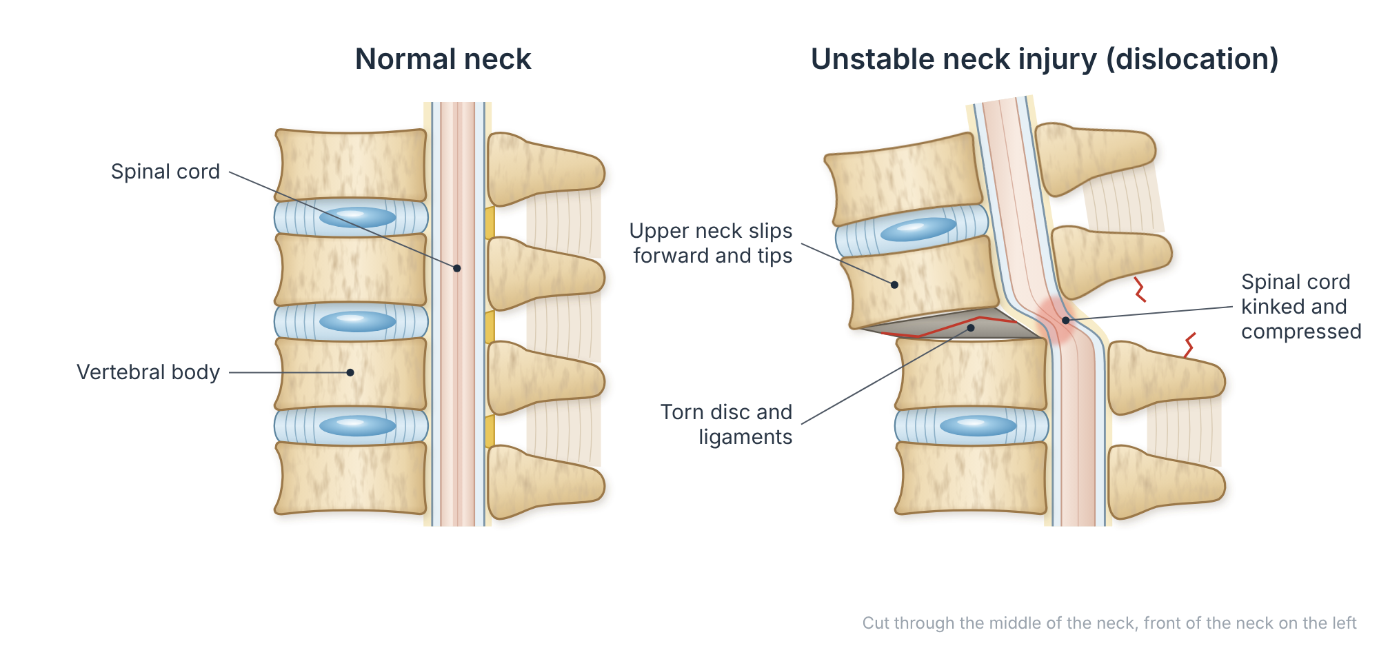
At a glance
- Two age groups: young people injured in high-energy accidents (road accidents, diving, rugby, falls from height), and older people who fracture the top of the neck in a simple fall.
- Upper neck (C1 and C2): the commonest fracture in older people is through the peg of C2 (odontoid or dens fracture).
- Lower neck (C3 to C7): injuries range from stable compression fractures to burst fractures and dislocated facet joints.
- Stable fractures usually heal in a firm collar over 6 to 12 weeks.
- Unstable fractures and those pressing on the spinal cord usually need surgery to realign, decompress and stabilise the neck.
Causes
In younger people, neck fractures usually follow high-energy injuries: motor vehicle and motorcycle accidents, diving into shallow water, contact sports such as rugby, and falls from a height. In older people with thinner bones and stiff, arthritic necks, a simple fall onto the face or head can be enough to break the upper neck.
Symptoms
- Neck pain after an injury, often severe and made worse by movement
- Tenderness over the back of the neck
- Pins and needles, numbness or weakness in the arms or legs if the nerves or spinal cord are affected
- Difficulty with balance, walking or bladder control in more severe spinal cord injuries
Emergency: protect the neck
After any significant injury, anyone with neck pain, numbness, tingling or weakness should be kept still and taken to an emergency unit. Do not move the head to “test” the neck. Early treatment of spinal cord compression gives the best chance of recovery.
How is it diagnosed?
Doctors use well-tested decision rules, such as the Canadian C-spine Rule, to decide who needs a scan. A CT scan is the best test to show fractures. An MRI scan is added when there are nerve symptoms or when damage to the ligaments, discs or spinal cord is suspected.
Types of neck fracture
Upper neck (C1 and C2)
- C1 (atlas) ring fractures: often stable and treated in a collar.
- C2 peg (odontoid) fractures: the commonest neck fracture in people over 70. Some heal in a collar; others do not and need surgery.
- “Hangman’s” fractures of C2: most heal in a collar or brace; displaced fractures may need fixation.
Lower neck (C3 to C7)
- Compression fractures: the front of the vertebra is squashed; usually stable.
- Burst fractures: the whole vertebral body breaks and fragments can press on the spinal cord.
- Facet dislocations and fracture-dislocations: one vertebra slides forward on the next, often injuring the spinal cord. These are unstable and need urgent realignment.
Non-surgical treatment
Stable fractures are treated in a firm, well-fitted cervical collar, usually for 6 to 12 weeks, with X-rays to check that the bones stay aligned while they heal. Pain relief and early walking are encouraged. Once the fracture has healed, gentle return to activity follows; non-impact cardio such as walking, cycling, spinning or swimming helps recovery.
Surgical options
Surgery is recommended when the neck is unstable, when bone or disc is pressing on the spinal cord or nerves, or when a fracture is unlikely to heal in a collar.
Anterior (front of the neck) surgery
Through a small incision at the front of the neck, the damaged disc or vertebra is removed, the spinal cord is freed, and the gap is filled with a cage or bone graft held by a plate and screws. This is commonly used for burst fractures and disc injuries.
Posterior (back of the neck) fixation
Screws placed into the vertebrae from behind are joined by rods to hold the neck in the corrected position. This is often used for dislocations and injuries of the ligaments at the back of the neck, sometimes combined with front surgery.
Upper neck surgery
Odontoid fractures can be fixed with a screw through the front of C2, or by fusing C1 to C2 from behind with screws and rods (see C1–C2 fusion).
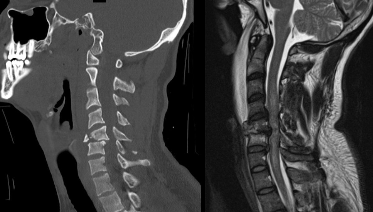
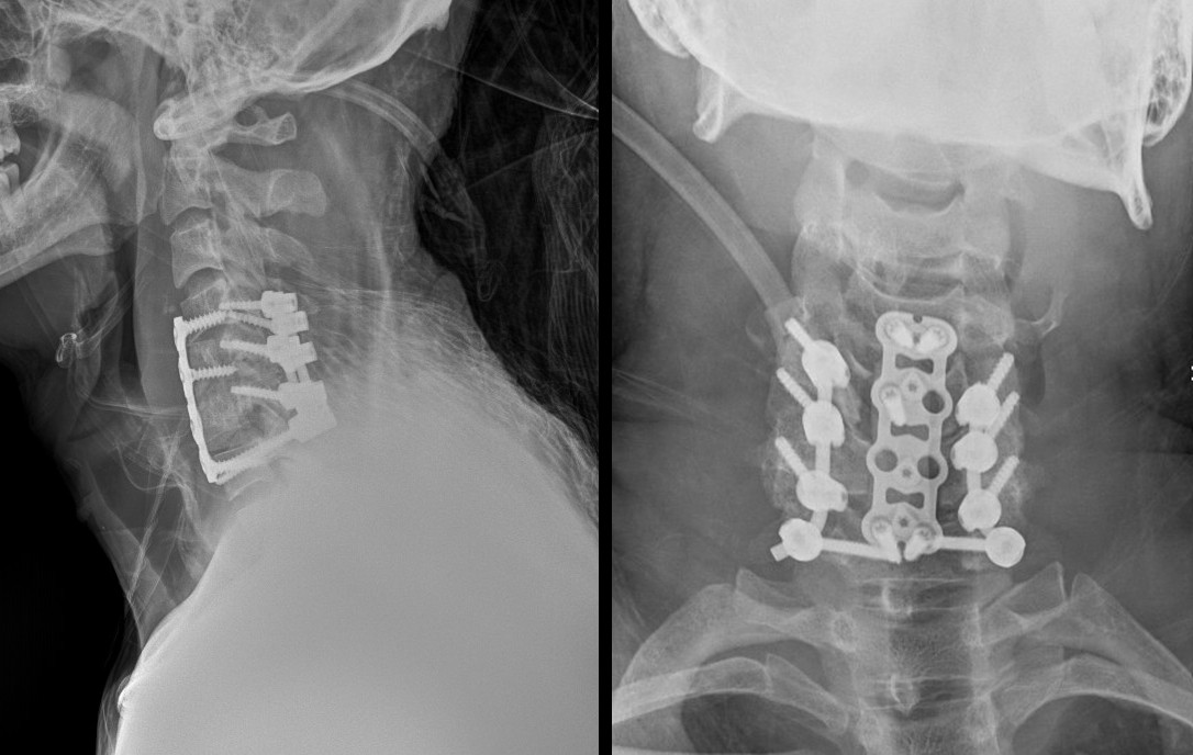
Example: C5/6 fracture-dislocation
This was a high-energy neck injury with a fracture-dislocation at the C5/6 level. The spinal cord was decompressed and the neck was reconstructed with a combined approach from the front and the back.
What the evidence says
In the STASCIS study of people with spinal cord injury in the neck, those whose spinal cord was decompressed within 24 hours of injury were more likely to recover meaningful neurological function than those operated on later. This is why spinal cord compression is treated as an emergency.
Recovery
Most people with stable fractures make a good recovery once the bone has healed. After surgery, most patients are up and walking within a day or two and wear a soft or firm collar for a few weeks. Recovery after a spinal cord injury depends on how severe the injury was, and is supported by specialised rehabilitation.
Talk to Prof Kruger
If you or a family member has this condition and would like an opinion on the options, you are welcome to book a consultation. Please bring any recent scans and reports.
References and key studies
- Stiell IG, et al. The Canadian C-spine rule for radiography in alert and stable trauma patients. JAMA. 2001;286(15):1841–1848.
- Vaccaro AR, et al. The subaxial cervical spine injury classification system: a novel approach to recognize the importance of morphology, neurology, and integrity of the disco-ligamentous complex. Spine. 2007;32(21):2365–2374.
- Vaccaro AR, et al. AOSpine subaxial cervical spine injury classification system. Eur Spine J. 2016;25(7):2173–2184.
- Fehlings MG, et al. Early versus delayed decompression for traumatic cervical spinal cord injury: results of the Surgical Timing in Acute Spinal Cord Injury Study (STASCIS). PLoS One. 2012;7(2):e32037.
- Vaccaro AR, et al. Functional and quality-of-life outcomes in geriatric patients with type-II dens fracture. J Bone Joint Surg Am. 2013;95(8):729–735.
This page is general patient education based on published research and is not a substitute for a personal consultation. Treatment is always tailored to the individual.